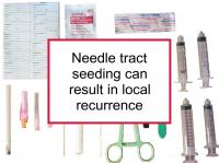
Needle tract seeding (also known as needle track seeding) occurs when breast cancer cells are deposited along the path of a biopsy needle as it is withdrawn from the breast. Evidence suggests that such seeding occurs 20% to 50% of the time. However, most breast cancer cells moved by biopsy needles do not survive — studies that have examined the question have been reassuring.
Nevertheless, cases have been described in which local recurrences were found in the biopsy path. Such "successful" malignant seeding has been estimated to occur approximately 0.07% of the time when surgery is followed by radiotherapy and 7% when radiation is omitted.
Now a new study has reported that high tumor grade, triple negative (ER-/PR-/HER2-) disease and multiple needle insertions appear to be risk factors for malignant needle tract seeding.
Needle tract seeding
The mechanical force of a needle biopsy can displace tumor cells. Biopsies also cause bleeding and other fluid movement that can disseminate tumor cells. Displaced tumor cells have been observed in the needle tracts in surgically removed tissue of women who previously underwent needle biopsies.
Needle tract seeding has also been demonstrated in a number of cases in which metastases arose along the needle tract. However, cases have also been reported of skin seeding and lymph node seeding, as well as increases in circulating tumor cells, after needle biopsies.
Skin seeding has the potential to result in skin metastasis. In one case, a women who underwent a multiple-puncture biopsy was found to have nests of cancer cells in the skin after the biopsy. She went on to have recurrence in the skin at the biopsy site after 34 months.
Isolated tumor cells have been shown to be more common in the sentinel lymph node after needle biopsy, indicating displacement of tumor cells. However, note that benign breast cells can also move into axillary lymph nodes after biopsy, which can result in a false positive lymph node status determination. Breast cancer surgeries and biopsies are known to result in increased concentrations of circulating tumor cells.
Avoiding metastasis from needle tract seeding
What can be done to minimize the possibility of breast cancer recurrence from needle tract seeding? As noted above, isolated tumor cells are more commonly found in the sentinel lymph node after breast cancer needle biopsy, indicating displacement of tumor cells beyond the needle tract. However, similar cancer cell displacement also appears to occur after surgical removal of a breast tumor. Therefore, having a surgical biopsy instead of a needle biopsy is not the answer.
It is preferable that the lumpectomy (or more extensive breast conserving surgery) that follows a biopsy remove all of the tissue immediately surrounding the biopsy path. In addition, radiation treatment appears to be effective in destroying breast cancer cells dispersed in the breast by needle biopsies.
However, the needle tract must be part of the radiation field. This is automatic in the case of whole breast irradiation, but not for partial breast irradiation, in which case the biopsy puncture site might be outside the field. These observations imply that the location of the entrance point of the needle (or needles) should be recorded. Recently, it has been suggested that treating the biopsy cancel by inserting a gelatin stick infused with a chemotherapy drug could reduce tumor cell seeding.
Latest research finds aggressive disease is a higher risk for malignant seeding
The study referenced at the beginning of this news story was designed to investigate factors associated with malignant seeding following image-guided needle biopsy of the breast. To conduct the study, the authors performed a retrospective review of 4,010 women would underwent biopsies for breast cancer or suspicious breast findings and subsequently were diagnosed with malignant needle tract seeding. Needle guidance method, needle gauge, and number of passes were noted. In addition, histology, tumor grade, hormone receptor and HER2 status, T category, and N category were documented.
Only eight cases of confirmed malignant needle tract seeding were found. The average time from biopsy to a diagnosis of malignant seeding was 61 days. Almost all (7/8) of the cases were classified as invasive ductal carcinoma. The majority (6/8) were grade 3 (75.0%). In addition, more than half (5/8) of the cases were triple negative. Almost all (7/8) of the women underwent biopsy with ultrasound guidance. Multiple-insertion, non-coaxial ultrasound-guided core-needle biopsy was performed in six cases. The malignant needle tract seeding was evident on mammogram as focal asymmetry (3/7 cases), mass (1/7), calcifications only (1/7), or not visible (occult) (2/7). Sonography showed a mass in almost all (7/8) cases, in many cases with irregular shape (5/7) and without circumscribed margins (6/7). No visible seeding was evident in one case. Seeding was found to be both intradermal (within the skin) and subdermal.
The authors conclude that high-grade, triple negative breast cancers and multiple-insertion, non-coaxial biopsies could increase the risk of malignant needle tract seeding. Such seeding should be suspected on the basis of the superficial and linear pattern of disease progression in these patients, according to the authors.
